Modern features of ultrasonic sensors
13 January 2022The ultrasound transducer is an essential attribute of ultrasound scanners. How to choose the right one
to choose an ultrasound transducer in accordance with the tasks to be performed to obtain
the highest quality image of the area of interest? Let's try to figure it out.
We will not describe in detail how ultrasound waves are generated and
are generated and the image is formed, because the physics of ultrasound is the basis of knowledge of every
specialist involved in ultrasound diagnostics.
Let's start with a brief classification. First of all, it is a classification by
constructive parameters:
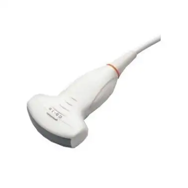
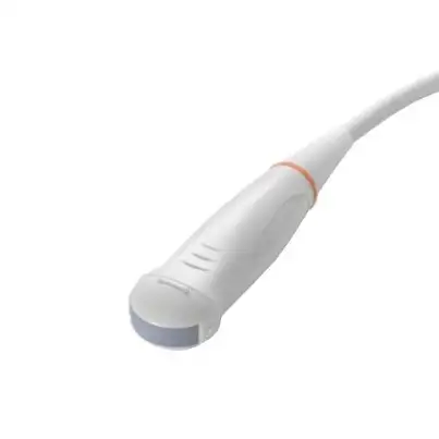
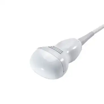
Convection sensor. Inuses a frequency of 2.5-7.5 MHz. It has a shorter length, so it is easier to achieve
uniformity of its adherence to the patient's skineasyand more. However, when
using convex sensors, an image is obtained with a width of several
centimetres wider than the size of the transducer itself. To clarify the anatomical
landmarks, the doctor must take this discrepancy into account. Due to the lower
frequency, the scanning depth reaches 20-25 cm. It is usually used for
examination of deeply located organs - abdominal and retroperitoneal organs
space, genitourinary system, hip joints.
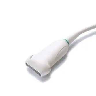
Linearand sensor. Вuseis
frequency of 5-15 MHz. The advantage of the linear sensor is the full compliance of the
of the organ under examination to the position of the sensor itself on the body surface. The disadvantage
of linear transducers is the difficulty of ensuring uniform
adherence of the transducer surface to the patient's skin in all cases, which leads to distortions
of the image at the edges. Also, linear sensors, due to their higher
frequency allows you to obtain images of the study area with high
resolution, but the scanning depth is quite small (no more than 10 cm, although manufacturers often write a depth
is much greater - this is not true). They are used mainly for the study of superficially
structures - thyroid gland, mammary glands, small joints and
muscles, as well as for the study of blood vessels.
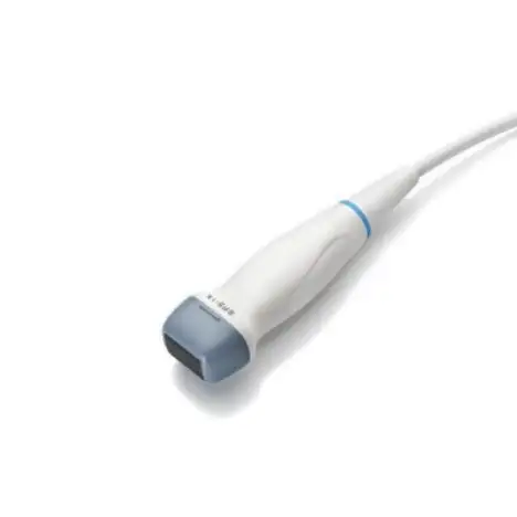
Phased/Sector Sensor. Poperates at a frequency of 1.5-5 MHz. It has an even greater discrepancy between
between the size of the sensor and the resulting image, so it is used mainly in
cases where it is necessary to get a large overview of a small area of the body at
depth from a small area of the body. The most appropriate use of sectoral scanning is in examinations,
for example, through intercostal spaces. A typical application of the sector transducer
is echocardiography - examination of the heart, examination of brain vessels (transcranial scanning),
examination of pleural cavities and lung tissue
Biplane sensor. It is a combination of two types of emitters
(convex+convex or convex+linear), which allows to obtain images in
longitudinal and transverse sections. There are even three-plane transducers, but
they have not gained wide popularity in ultrasound diagnostics.
Biplane transducers are currently used mainly in urology to assess
prostate gland
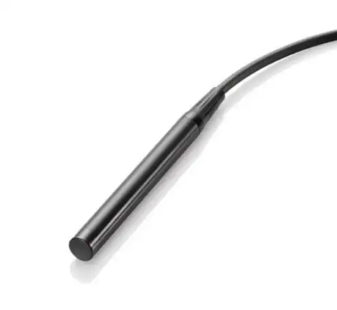
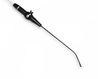
Pencil sensor (blind CW). Pencil Doppler sensors are used for
examination of the great vessels of the extremities and neck with a frequency of 2-8 MHz using
constant-wave Doppler (CW Doppler). Transducers containing a separate
emitter and receiver - can operate in B-mode and DCE. However, due to the fact that
with the fact that the use of CW Doppler in modern ultrasound scanners is now possible on all
transducers, the expediency of using pencil transducers has lost its relevance.
At present, given the low price of these transducers, they are still manufactured by
manufacturers and are still used in medical practice
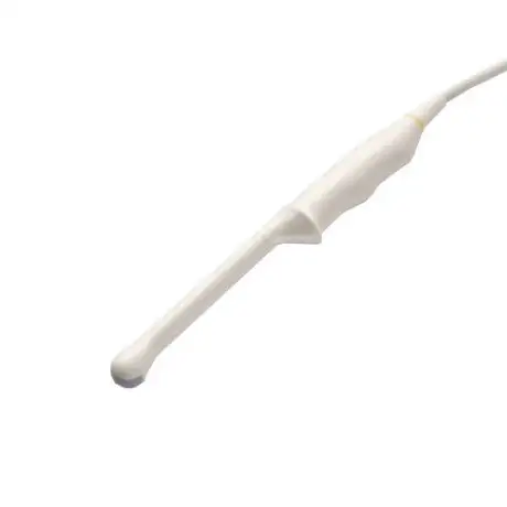
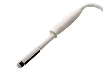
Intracavitary sensor. In turn, they are divided into vaginal,
rectal, vaginal-rectal, transurethral. The scanning surface can be
represented by convection, phase or ruler. The viewing angle and frequency of such
of such sensors are quite diverse. There is a wide range of intracavitary
intracavity transducers directly for biopsies, the biopsy needle passes directly
through the body of the transducer itself. The most popular among clinicians
intracavity transducer is a transducer with a convex scanning head, with a
scanning frequency of 5-9 MHz, viewing angle - from 90° to 180°
Modifications of traditional gauges:
 Microconvection Sensors. This is a convection sensor used in
 Peresophageal transducers. They are used for echocardiography.
There are paediatric and adult transducers with different lengths and diameters. Operating frequency is 4-9 MHz,
the angle is about 90°. Modern variations suggest a bi-planar structure, but
most often these are sectoral and microconvection sensors.
Peresophageal transducers. They are used for echocardiography.
There are paediatric and adult transducers with different lengths and diameters. Operating frequency is 4-9 MHz,
the angle is about 90°. Modern variations suggest a bi-planar structure, but
most often these are sectoral and microconvection sensors.
 Laparoscopic/intraoperative sensors. They are used in laparoscopic
interventions. Their feature is the ability to control with a joystick. Intraoperative
ultrasound sensors of the device are designed to visualise the surgeon's actions in the surgical field.
Laparoscopic/intraoperative sensors. They are used in laparoscopic
interventions. Their feature is the ability to control with a joystick. Intraoperative
ultrasound sensors of the device are designed to visualise the surgeon's actions in the surgical field.
 Volumetric (3D/4D) convex or linear sensor. Scanning is performed by
using a scanning head driven by a motor located in the sensor itself.
sensor itself. It is used to obtain three-dimensional images. It is most widely used in
Volumetric (3D/4D) convex or linear sensor. Scanning is performed by
using a scanning head driven by a motor located in the sensor itself.
sensor itself. It is used to obtain three-dimensional images. It is most widely used in
The next important parameter of ultrasonic sensors is the technology
of production. According to the technologies used in ultrasonic sensors,
are:
Piezocrystalline (standard) sensors. The most popular for use in
clinical practice due to their relatively low cost and widespread production. The more
more densely the elements are arranged, the greater the resolution we can
we can obtain. For example, convex and linear sensors of standard density contain
128 elements, while similar high-density emitters contain 192 elements. However,
it is not the number of elements that is important. What matters is the density of the lines, which
depends on the geometric parameters of the sensors (aperture, viewing angle, radius).
Single-crystal sensors. The difference in the technological process is
that standard piezocrystalline sensors are assembled from individual
piezoelectric elements. Although they are slightly different, this has a negative impact on
the quality of the image. In single-crystal sensors, piezoelectric elements are "sliced"
from a single crystal. The image obtained on such sensors is less "noisy" (more
cleaner), even before any computer processing of the signal.
Matrix sensors. In classical ultrasonic emitters, piezoelectric elements (128-256) are arranged
in one row. Matrix transducers have from 3 to 10 such rows - a matrix. Several
rows of piezoelectric elements form a beam that is thinner and more uniform in
in thickness. The narrower the beam, the higher the image resolution. This effect is achieved by
high-density sensors, but only at a certain depth (in focus). Matrix sensors
sensors, equally well visualise the near, middle and far scanning area.
Such emitters are 2 times more expensive than classical ones. Depending on the number of
rows, there are 1.5D and 2D (when the number of elements is almost the same on both
sides and a three-dimensional image can be obtained).
The final step when choosing an ultrasonic sensor is to pay attention to
geometrical and acoustic parameters -
aperture (working surface), radius of curvature and viewing angle - respectively.
If we conditionally divide ultrasound scanners of all available manufacturers into 3
groups: basic (low-end devices), middle class and expert ultrasound
scanners, it is worth noting that the number of supported transducers is directly proportional to the
increases with the class of the device. Thus, basic devices are limited to 1-2 standard sensors
for the main types of examination.
class devices have a fairly wide range of sensors with different frequencies and support
and support single-crystal technology. Experts, as a rule, have more than
dozens of sensors in their arsenal, which will satisfy even the most demanding clinician,
manufacturers produce matrix sensors exclusively for "experts". Considering
these features, when choosing a transducer, try to get the most out of the ultrasound machine
machine and get the highest quality and clearest image that will not give the
the doctor to make a diagnostic mistake.
Author: Dmytro Yakovenko, ultrasound doctor, applicant of Harwind
-
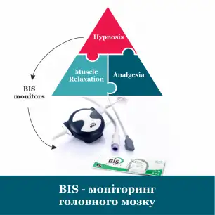
![Article]() BIS - brain monitoring21 July 2023
BIS - brain monitoring21 July 2023 -
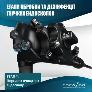
![Article]() Stages of processing and disinfection of flexible endoscopes. Stage 1: primary cleaning of the endoscope.27 May 2022This stage is carried out in the treatment room immediately after the examination, before disconnecting the device from the illuminator, aspirator and video processor. It is very important to clean and rinse the endoscope, inside and out, immediately after the procedure to prevent contaminants from drying and sealing.
Stages of processing and disinfection of flexible endoscopes. Stage 1: primary cleaning of the endoscope.27 May 2022This stage is carried out in the treatment room immediately after the examination, before disconnecting the device from the illuminator, aspirator and video processor. It is very important to clean and rinse the endoscope, inside and out, immediately after the procedure to prevent contaminants from drying and sealing.
 The basket is empty